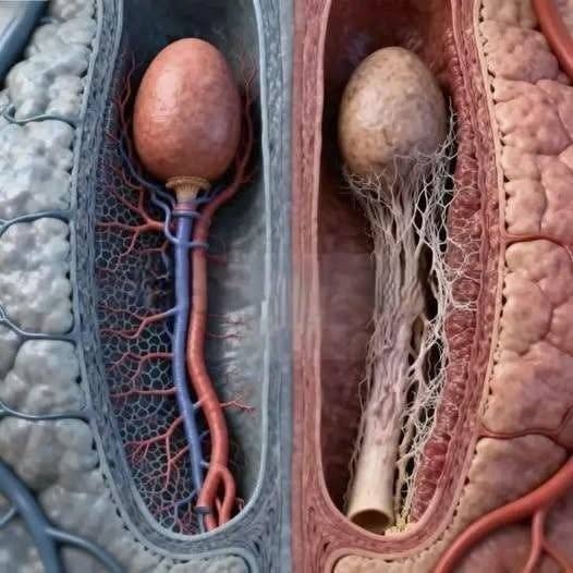
Digital media platforms frequently feature sensationalized, pseudo-anatomical computer graphics paired with incomplete clickbait headlines—such as claiming that elderly men undergo drastic, unnatural internal anatomical transformations. These viral posts are specifically engineered to provoke curiosity, health anxiety, and rapid online comment sharing rather than providing accurate, evidence-based medical or anatomical education.
From a clinical anatomy and urological perspective, the rendered image does not depict real human tissue or documented anatomical pathology. Instead, it is a synthetic 3D rendering that visually mimics biological structures—often repurposed from botanical illustrations, abstract digital art, or artificial intelligence models—to falsely represent blood vessels and internal organs. While the male reproductive system and vascular network undergo natural physiological changes with age (such as benign prostatic hyperplasia or microvascular stiffness), human anatomy does not develop bizarre, isolated plant-like roots or structural metamorphoses as depicted in clickbait graphics. Sensationalized online content regularly uses fabricated visuals to manufacture intrigue and maximize digital engagement metrics.
Understanding human aging and anatomical health relies on verified medical literature, clinical imaging (such as ultrasound, MRI, or CT scans), and professional healthcare guidance rather than unverified digital renders. Personal health decisions and biological understanding should always reflect established clinical science. If you have questions regarding age-related physiological changes, vascular health, or urological wellness, consulting a licensed physician or specialist provides safe, scientifically sound direction.
Leave a Reply